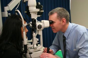
Comprehensive ocular examinations are imperative for the continued health of the eyes. It includes a prescription check, cataract assessment, glaucoma evaluation and pressure check after which a plan is suggested which pertains to your individual needs. Both Darryl and Maria are therapeutically qualified so are able to provide further ocular health management and advice. Should ophthalmologic evaluation be required, this can also be arranged.
Children should have an eye exam by no later than 6 months of age, then again by age 3, and just before starting school. School-age children need an exam every two to three years after that if they have no visual problems. If your child requires eyeglasses or contact lenses, schedule visits every 12 months.
Prescriptions change frequently, because vision matures along with your child. Your optometrist will also ensure that your child has the visual skills necessary for succeeding in school, such as using the eyes as a team, peripherial vision, ease of focusing from distance to near and eye/hand coordination.
Glaucoma is the name given to a group of related diseases where the optic nerve is being damaged. The nerve fibres progressively die taking away the peripheral or side vision first. Visual loss often goes undetected until it is quite advanced. Glaucoma is the number one cause of preventable blindness in New Zealand and other developed countries.
Glaucoma often has no symptoms, so regular eye exams are essential. Elevated eye pressure is an important risk factor for developing glaucoma. Treatment for glaucoma is determined by a variety of factors but aimed at lowering pressure to prevent further loss of vision.
At Wight we not only diagnose it but we are specialists in co-managing it, even up to treating it.
A cataract is a clouding of the eye’s natural lens, which lies behind the iris and the pupil. The lens is mostly made of water and protein. As we age, some of the protein may clump together and start to cloud a small area of the lens. This is a cataract, and over time, it may grow larger and cloud more of the lens, making it harder to see.
There are three types of cataracts, sub capsular, nuclear and cortical .
Various cause of cataracts include, exposure to ultraviolet light, diabetes, use of steroids, diuretics and major tranquilizers, cigarette smoke, air pollution and heavy alcohol consumption. A diet high in antioxidants , such as beta-carotene (vitamin A), selenium and vitamins C and E, may forestall cataract development.
Initially vision may be able to be improved for a while using new spectacles, higher magnification, appropriate lighting or other visual aids. However cataract surgery is usually the eventual treatment. Cataract surgery is very successful in restoring vision. In fact, it is the most frequently performed ocular surgery in the New Zealand. Nine out of ten people who have cataract surgery regain very good vision, somewhere between 6/6 and 6/12.
Both Maria and Darryl are therapeutically qualified to diagnose and treat most anterior eye conditions and write prescriptions should that be needed. Conditions such as conjunctivitis, blepharitis, red eye, itchy eye, watery eyes, dry eyes, uveitis and post-cataract complications can all be managed at Wight Optometrists.
A few years ago the face of Optometry changed in New Zealand whereby certain optometrists after further intense post graduate studies were given the privilege to write prescriptions and manage anterior eye pathology. Both Maria and Darryl took up the challenge and have been practising as therapeutic optometrists for the last three and a half years.
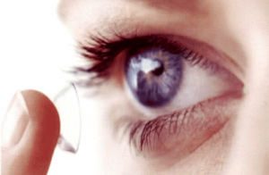
A contact lens fitting examination includes a full general vision assessment, a prescription check and health evaluation. The most appropriate and individually tailored contact lens option is then suggested after which suitable lenses are fitted and assessed. The contact lens wear, care and management is then taught and the process continues.
Disposable soft contact lenses, coloured lenses, extended wear contacts and rigid gas permeable lenses are fitted on site.
Keratoconus is a hereditary corneal condition which is progressive and visually compromising. There is often a high degree of irregular astigmatism which compromises general vision. Keratoconus requires professional eye care and often rigid gas permeable lens fitting, both of which are a significant part of this practice.
In order to determine if you are suitable to for laser vision surgery an assessment and individually tailored advice is necessary. This assessment involves comparing your previous ocular refraction to your current prescription, an ocular health check and evaluating your corneal integrity. You shall then be advised accordingly and reputed laser vision specialists shall be recommended.
Both level 1 and level 2 visual examinations are possible at Visique Wight Optometrists. You need to present a visual form from the Police force. The assessment usually takes about forty five minutes.
With a family community services card, a child under the age of 16 is granted a subsidy towards an eye examination and spectacles every year. A current valid community services needs to be presented at the eye examination.
Please contact us to discuss the details of this subsidy and how it could benefit your child.
A government subsidy exists for those who require contact lenses to aid with high degrees of short-sightedness and keratoconus. This subsidy is supported by Visique Wight Optometrists and requires a comprehensive visual test and professional expertise.
This examination usually takes about twenty minutes and will be suggested by Maria or Darryl should a more extensive, objective assessment be required to provide information of your ocular health and perhaps general wellbeing. Visual field loss may occur due to disease or disorders of the eye, optic nerve, or brain. A general routine visual field assessment is often advised after the age of forty to provide a baseline result for future reference.
In order to enable a more thorough examination of the retina and peripheral ocular structures, dilating drops are used to widen the pupils to ensure a more comprehensive and holistic view is gained.
We have a state of the art digital fundus camera which enables us to take clear and accurate pictures of the retina, optic nerve and blood vessels. This is valuable in monitoring the progress of conditions such as diabetic retinopathy, glaucoma, macular degeneration and ocular fundi lesions. It is one of the most accurate ways to illustrate and view the peripheral fundus.


